tree in bud radiology assistant
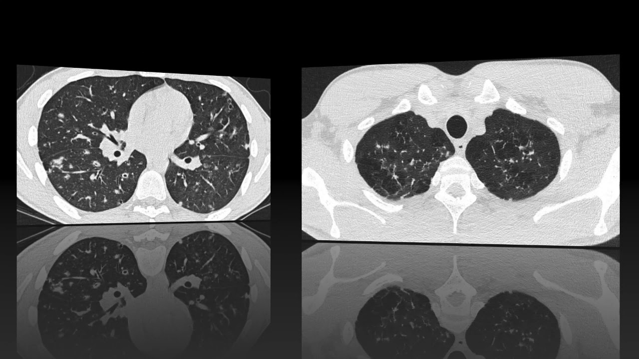
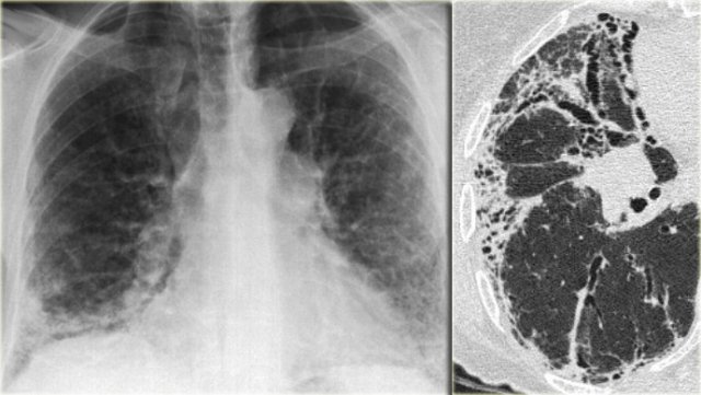
Upper and middle zone predominance. CTHRCT high-resolution-CT showed extensive bronchiectasis along with parenchymal disruption in the right upper lobe.
Chronic Airspace Disease Review Of The Causes And Key Computed Tomography Findings
We aimed to establish the incidence of the TIB pattern as a proportion of all patients undergoing chest CT.
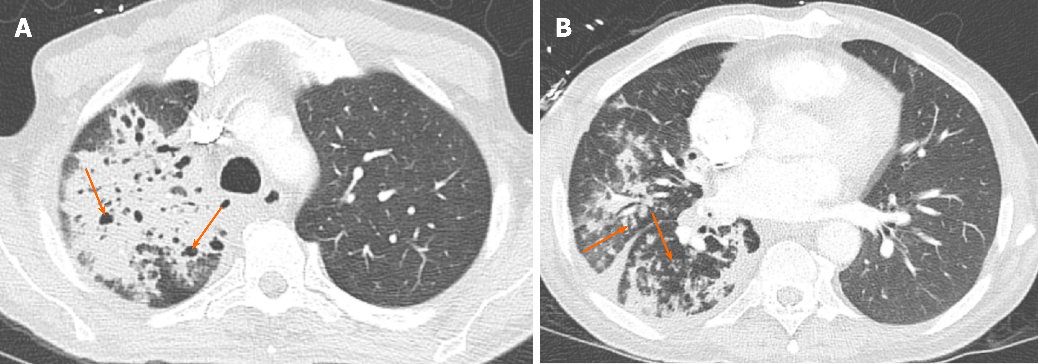
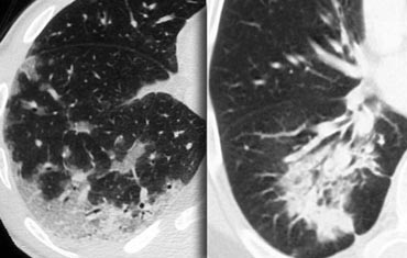
. 78 indicating the absenceresolution of TIB opacities 26 incomplete thoracic CT scan studies 75 duplicate. Thin section CT shows peribronchial thickening and centrilobular nodules with tree in bud appearance. Tree-in-bud TIB is a radiologic pattern seen on high-resolution chest CT reflecting bronchiolar mucoid impaction occasionally with additional involvement of adjacent alveoli.
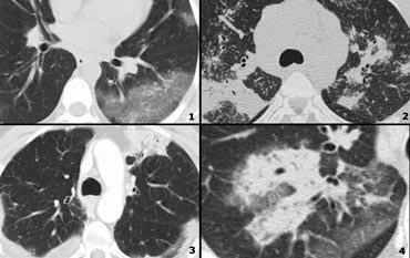
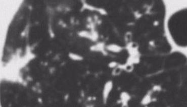
It consists of small centrilobular nodules of soft-tissue attenuation connected to multiple branching linear structures of similar caliber that originate from a single stalk. The tree-in-bud appearance characterised by well-defined centrilobular nodules was observed in 1 29 patient. Small nodules some related to vessels admixed with ground-glass opacity and consolidation.
Small nodules in a perilymphatic distribution ie. Tree in bud opacification refers to a sign on chest CT where small centrilobular nodules and corresponding small branches simulate the appearance of the end of a branch belonging to a tree that is in bud. ECR 2014 C-1769 Classic Signs in Thoracic Radiology by.
Abnormal tree-in-bud bronchioles can be distinguished from normal centrilobular bronchioles by their more irregular appearance lack of tapering or knobbybulbous appearance at the tip of their branches. Dishes are a symbol of a familys welfare. Of Medicine Medical College 88 College Street Kolkata 700 073.
Originally reported in cases of endobronchial spread of Mycobacterium tuberculosis this. The tree-in-bud appearance may occur in case of distal airway diseases in bacterial viral and fungal infections in some congenital diseases for example cystic fibrosis in some idiopathic. CT tree-in-bud is most commonly reported in peripheral airways disease related to an infectious etiology.
Received November 11 1999. Pus mucus or inflammatory exudate centrilobular bronchioles. It represents dilated and impacted mucus or pus-filled centrilobular bronchioles.
Revision requested December 10. Tree-in-bud describes the appearance of an irregular and often nodular branching structure most easily identified in the lung periphery. Along subpleural surface and fissures along interlobular septa and the peribronchovascular bundle.
In centrilobular nodules the recognition of tree-in-bud is of value for narrowing the differential diagnosis. The Tree-in-Bud Sign. 1 From the Department of Radiology University of Vienna Waehringer Guertel 18-20 A-1090 Vienna Austria.
Endobronchial spread of infection TB MAC any bacterial bronchopneumonia Airway disease associated with infection cystic fibrosis bronchiectasis less often an airway disease associated primarily with mucus retention allergic bronchopulmonary aspergillosis asthmaDiagnosis Radiology. 657 and bronchial wall thickening and consolidation n22. Lymphadenopathy in left hilus right hilus and paratracheal 1-2-3 sign.
Tree-in-bud pattern and poorly defined nodules representing bronchiolar filling. Mar 25 2020 - Poster. To make it even worse it was bordering on my neighbor s lot and the old man who lived there was even more superstitious than most.
3 found that the tree-in-bud pattern was seen in 256 of the CT scans in patients with bronchiectasis. She also had generalized weakness and shortness of breath for same duration. Based on lesion localization and presence or suspicion of a concomitant bronchial disease for cats in this sample authors propose that the CT treeinbud pattern described in.
Its microbiologic significance has not been systematically evaluated. Primary pulmonary lymphoma or leukemia 247. Our Radiology Information System was searched for the term tree-in-bud from January 1 2010 to December 31 2010 iden-tifying 599 examinations.
In conclusion the treeinbud pattern should be considered as a differential diagnosis for radiographic soft tissue opaque nodules in feline lungs. Trees in front of the inclined houses. Of these 182 cases were excluded for the following reasons.
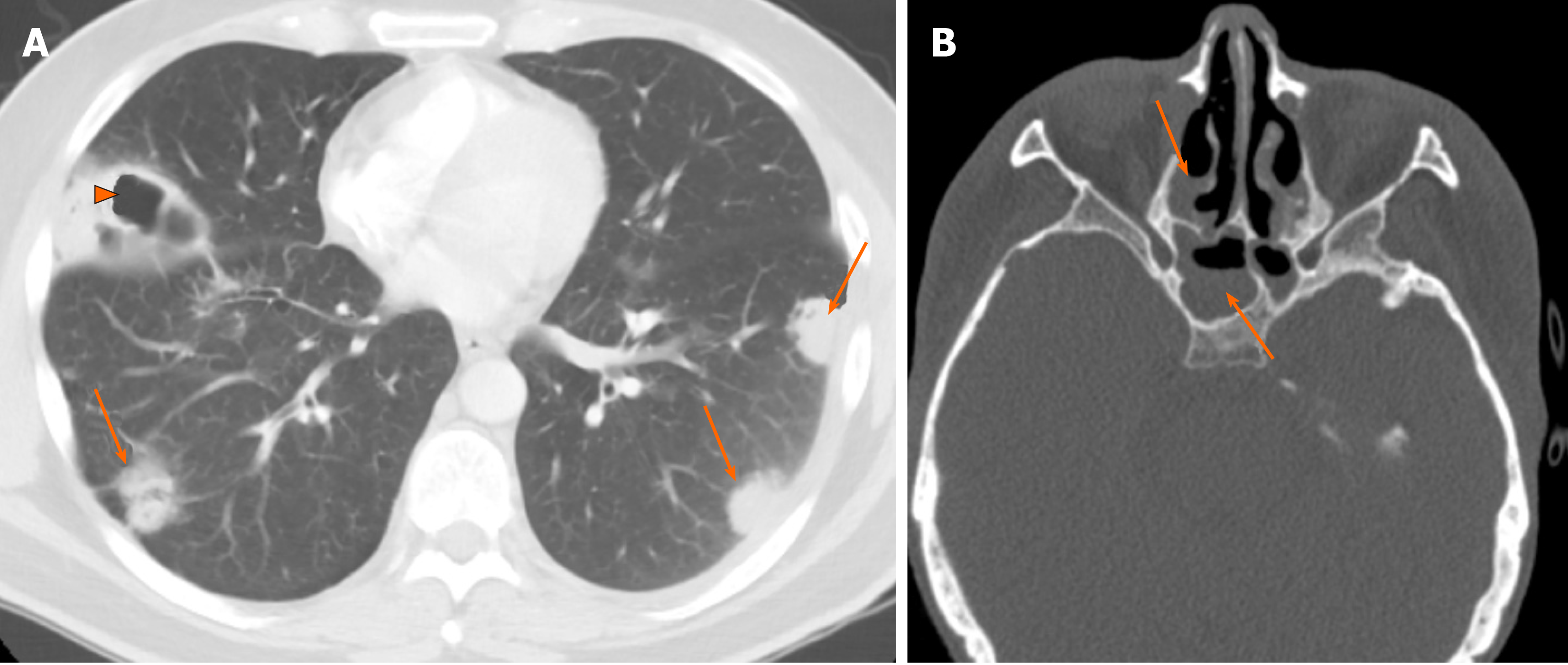
The tree-in-bud pattern is commonly seen at thin-section computed tomography CT of the lungs. Tree-in-bud almost always indicates the presence of. The patient had an oesophageal lesion below the carina extending longitudinally 6 cm.
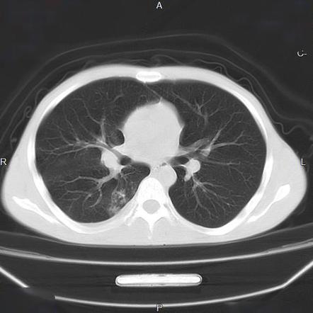
Despite atypical findings for COVID-19 pneumonia RT-PCR test was positive for COVID-19. The remaining pulmonary parenchyma demonstrated scattered tree-in-bud pattern with lower lobe predominance and without pleural effusion. Tree-in-bud appearance represents dilated and fluid-filled ie.
Differential diagnosis is broad which includes different etiologies. Revision received and accepted May 22 2000. Tree exactly opposite to the southwest entrance door is not at all menace.
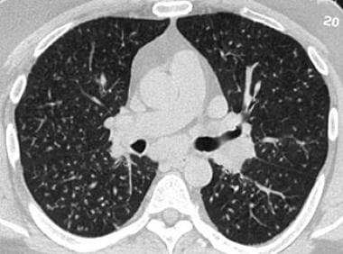
886 followed by GGA and consolidation n23. Usually somewhat nodular in appearance the tree-in-bud pattern is generally most pronounced in the lung periphery and associated with abnormalities of the. 1 refers to a pattern seen on thin-section chest CT in which centrilobular bronchial dilatation and filling by mucus pus or fluid resembles a budding tree Fig.
Tree in bud radiology assistant. Typical findings of BAC on HRCT include a solitary nodule or mass 43 focal or diffuse consolidation 30 or. Address correspondence to the author e-mail.
31 March 2013. A 50 years old lady presented with fever productive cough and occasional haemoptysis for two weeks. Originally and still often thought to be specific to endobronchial Tb the sign is actually non-specific and is the.
Another important entity that can produce the tree-in-bud pattern is bronchioalveolar carcinoma BAC 1. The most frequently observed combination of abnormalities was GGA and bronchial wall thickening n31. The identification of pulmonary vascular tree-in-bud in COVID-19 is unique and interesting.
Tree-in-bud pattern simulating diffuse panbronchiolitis but without cylindrical bronchiectasis.

Tree In Bud In Centrilobular Nodules The Recognition Grepmed

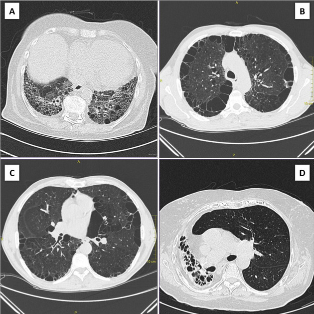
A Case Of Myelofibrosis With Anc Of 200 µl Hrct Chest Shows Multiple Download Scientific Diagram
2

Tree In Bud Sign Lung Radiology Reference Article Radiopaedia Org
The Radiology Assistant Hrct Basic Interpretation
Tree In Bud Sign Lung Radiology Reference Article Radiopaedia Org
The Radiology Assistant Hrct Common Diagnoses
The Radiology Assistant Hrct Basic Interpretation

Tree In Bud Almost Always Indicates The Presence Of Endobronchial Grepmed

Learningradiology Lung Abscess Pulmonary Lunges Pulmonary X Ray
Tree In Bud Sign Lung Radiology Reference Article Radiopaedia Org
Postprimary Tuberculosis Lung Imaging
The Radiology Assistant Hrct Common Diagnoses
Chronic Airspace Disease Review Of The Causes And Key Computed Tomography Findings
The Radiology Assistant Hrct Basic Interpretation
The Radiology Assistant Hrct Basic Interpretation
Honeycombing Lungs Radiology Reference Article Radiopaedia Org

Tree In Bud Pattern
2